VISION-DMD

Designed to ensure a timely & cost-effective drug development for Duchenne muscular dystrophy

Search

  • The Project
    • Summary
    • Project Objectives
    • Project Consortium
    • Contact us
  • About DMD
    • What is DMD?
    • Living with Duchenne
    • Current treatment options
    • Duchenne boy’s stories
    • DMD Community
    • Standards of Care for Duchenne muscular dystrophy
  • Study Information
    • What is a clinical study?
    • Phase 2a clinical trial information
    • Phase 2a preliminary study results
    • Phase 2b clinical trial information
    • Phase 2b Preliminary Results
    • MRI – Magnetic Resonance Imaging
  • Drug Development
    • Development of Vamorolone
    • Vamorolone development programme
  • Innovations
    • Return of patient data
    • Bone Health Outcomes
    • Biomarkers
    • Vamorolone study APP for boys
    • Patient Group Funders
    • What is Venture Philanthropy?
  • Resources
    • Relevant publications
      • VISION-DMD INFORMATION APP OVERVIEW
      • VISION-DMD Thought paper
    • VISION-DMD Workshops
      • Workshop series: Standards of Care
      • Workshop series: Biomarkers and Imaging
    • Webinars and presentations
    • Press Releases
    • VISION-DMD project Interviews
      • Collaborating for progress on rare diseases: an interview with Dr. Daria Julkowska, Scientific Coordinator of the European Joint Program on Rare Diseases.
      • Returning vamorolone study data to patient caregivers. What, when and how clinical trial data is returned to study participants. An interview with Dr. Laurie Conklin
      • An Interview with Andrea D’Alessandro – Clinical research project manager at TRiNDS
      • Interview with Jana Haberlová – Paediatric Neurologist at University Hospital Motol
      • Interview with Alex Johnson: a parent and patient foundation funder
    • VISION-DMD Project Newsletters
      • VISION-DMD newsletter sign up
    • VISION-DMD Poster presentations
    • Our privacy policy
  • English
    • Nederlands
    • Français
    • Deutsch
    • Italiano
    • Español

MRI – Magnetic Resonance Imaging

VISION-DMD investigated using quantitative Magnetic Resonance Imaging (MRI) to measure the difference in muscle involvement  in the Phase 2b study.

MRI is a technique able to produce pictures from inside the body. It is a completely painless and harmless technique. The child lies on a bed and their legs are moved to the centre of the tunnel. Their head remains near the scanner opening and it is possible for a parent or carer to stay in the room near them for reassurance.

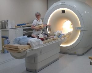
The child lies on the bed and enters the scanner feet first but their head stays out side the scanner bore. They are looked after throughout by a friendly radiographer! They are able to listen to stories or music during their scan. But be sure to keep nice and still!

Using MRI we can find out about the condition of the skeletal muscle in boys with Duchenne muscular dystrophy, and we can measure how it changes over time. In particular, we can find out how whether novel therapies can decrease the inflammation that is naturally present in the muscles of young boys with DMD as a measure of the acute disease process. Over time, the healthy muscle tissue is permanently replaced by fatty tissue and special MRI techniques are capable of measuring this very sensitively. By using MRI several times, we can estimate the effect that a therapy is having an effect on the underlying destructive processes.

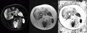
MRI is capable of distinguishing between the muscle tissue in the thigh that remains (left) and that which has been replaced by fat (middle) in Duchenne muscular dystrophy. From this information, MRI is capable of accurately measuring the percentage of muscle that has been replaced, and the change can be measured over time.

There have been a number of academic studies and trials which have used MRI as an endpoint. Sometimes the scanning sessions have lasted as much as an hour to collect comprehensive skeletal muscle data. Some boys find it difficult to keep still for this long. In this project, we are using our previous experience of DMD to present a simplified protocol which only requires 10 minutes of scanning (so the time in the scanner should be about 15-20 minutes in the scanner for boys who lie nice and still!).  Cutting down the patient burden is crucial for making the regular use of MRI a practical reality in DMD trials carried out in multiple centres.

Read our most recent publication

Follow us on twitter

  • Twitter

EUFlag2020

This project has received funding from the European Union’s Horizon 2020 research and innovation programme under grant agreement number 667078.

To find out more about the project contact us at [email protected]

VISION-DMD Copyright © 2023 · Log in

Multilingual WordPress with WPML